
Dog Knee Anatomy - The knee joint on a dog can be found in the hind leg, between the thigh and the lower leg.
Dog Knee Anatomy - In dogs, the most common knee injury is a rupture or tear of the cranial cruciate ligament. If you want to manage a knee injury, you might have a good piece of knowledge on the dog knee anatomy. If this plane were in the midline of the body, this is the median plane or median sagittal plane. • the dorsal plane divides the dog into ventral and dorsal portions. We help you understand its different parts and how they help your dog move freely.
You can guess what is the terminology while is watching the animation. Illustration of the anatomy of the dog’s knee. Cranial cruciate ligament (blue/purple), meniscus (red), caudal cruciate ligament (green). Positional and directional terms, general terminology and anatomical orientation are. Anatomy atlas of the canine general anatomy: There are two long bones, the femur (thigh bone) and the tibia (shin bone), and a small bone, the patella, which articulate together. The type of joint formed determines the degree and direction of motion.

canine knee anatomy
The mri of a normal left stifle of a dog was performed on a 1.5t mri by dr. Here, i will show you everything on the dog knee, including the bone involvement, ligaments, tendons and their arrangement with a labeled diagram. Main bone in lower leg. There are two long bones, the femur (thigh bone).

Canine Osteoarthritis Knee Model, Normal + 3 Conditions 1019577
Learn about the anatomy of your dog's knee. Muscle, organ and skeletal anatomy). The knee joint on a dog can be found in the hind leg, between the thigh and the lower leg. Connects to the hip bone via the hip joint. Learn about the anatomy of your dog's knee. Fully labeled photographs illustrating the.
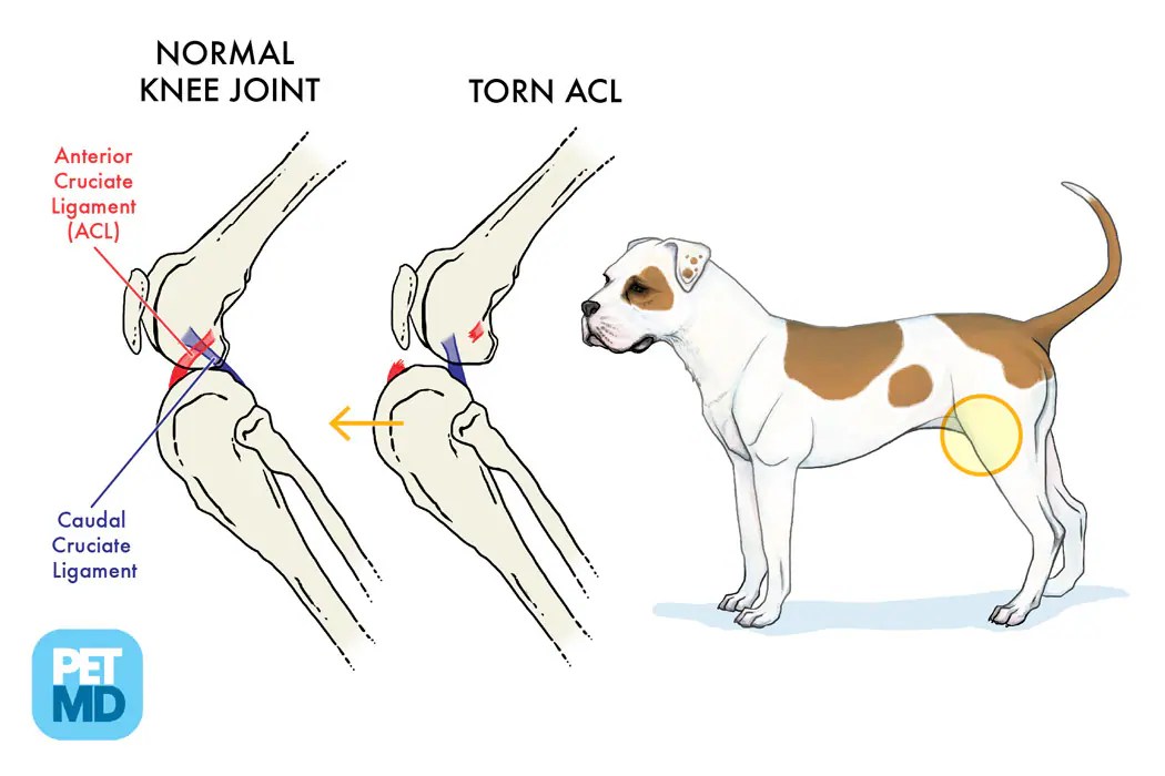
Cranial Cruciate Ligament Medical Diagram Torn Knee Ligament in Dogs
Understanding the location of the knee on a dog is essential for pet owners, veterinarians, and anyone interested in canine anatomy. Illustration of the anatomy of the dog’s knee. Anatomy atlas of the thigh, knee joint and leg in medial view. The knee is also called the stifle joint, which connects the tibia and fibula.

Canine Knee Anatomical Model 4Stage Osteoarthritis
Learn about the anatomy of your dog's knee. There are two long bones, the femur (thigh bone) and the tibia (shin bone), and a small bone, the patella, which articulate together. Dog knee and knee cap. If this plane were in the midline of the body, this is the median plane or median sagittal plane..

GPI 9050 Canine Knee Model
There are two long bones, the femur (thigh bone) and the tibia (shin bone), and a small bone, the patella, which articulate together. An overview of the key anatomy of the canine knee, including why structures are present, their clinical significance, and associated ligaments. Anatomy atlas of the canine general anatomy: Understanding the dog knee.

anatomy of the canine knee
The knee joint on a dog can be found in the hind leg, between the thigh and the lower leg. We help you understand its different parts and how they help your dog move freely. In dogs, the most common knee injury is a rupture or tear of the cranial cruciate ligament. You can guess.

Anatomical ModelCanine Knee
The dog stifle (knee) is anatomically very similar to a human knee. We’re putting dog leg anatomy into human terms to simplify and help you remember. The knee is also called the stifle joint, which connects the tibia and fibula with the patella, the dog version of a knee cap. Connects to the hip bone.

Anatomy of the canine (dog's) knee joint colorful design, healthy joint
Some joints do not move at all. While there are multiple ligaments within the knee, typically a torn knee ligament refers to the tearing of the cranial cruciate ligament. Connects to the hip bone via the hip joint. Dog knee and knee cap. Technically, the dog knee is on the rear legs. The knee joint.

ANATOMY OF THE DOG´S KNEE YouTube
Here, i will show you everything on the dog knee, including the bone involvement, ligaments, tendons and their arrangement with a labeled diagram. • the dorsal plane divides the dog into ventral and dorsal portions. Understanding the dog knee anatomy. Femur (thigh bone) patella (kneecap) tibia (shinbone) ligaments. Positional and directional terms, general terminology and.

Hills Pet Nutrition Dog Stifle Diagram patella, quadriceps, tibia
If you want to manage a knee injury, you might have a good piece of knowledge on the dog knee anatomy. Bones come together to form joints. Main bone in lower leg. Here, i will show you everything on the dog knee, including the bone involvement, ligaments, tendons and their arrangement with a labeled diagram..
Dog Knee Anatomy Dog leg anatomy is complex, especially dog knees, which are found on the hind legs. Anatomy atlas of the canine general anatomy: Small bone beside the tibia. Fully labeled photographs illustrating the dissection and the surgical approach of the medial femorotibial joint, with particular reference to the tibial plateau leveling osteotomy (tplo). Understanding the location of the knee on a dog is essential for pet owners, veterinarians, and anyone interested in canine anatomy.
Cranial Cruciate Ligament (Blue/Purple), Meniscus (Red), Caudal Cruciate Ligament (Green).
Main bone in lower leg. • the dorsal plane divides the dog into ventral and dorsal portions. Anatomy atlas of the canine general anatomy: • the sagittal plane divides the dog into right and left portions.
The Dog Stifle (Knee) Is Anatomically Very Similar To A Human Knee.
Anatomy atlas of the thigh, knee joint and leg in medial view. Illustration of the anatomy of the dog’s knee. If you want to manage a knee injury, you might have a good piece of knowledge on the dog knee anatomy. There are two long bones, the femur (thigh bone) and the tibia (shin bone), and a small bone, the patella, which articulate together.
The Insert Shows A Ruptured Cranial Cruciate Ligament (Also Note That The Shin Bone Is Displaced Forward And Crushing The Meniscus.
The type of joint formed determines the degree and direction of motion. It is a simple animation where i put one by one of the terminology of the dog´s knee. Connects to the hip bone via the hip joint. The mri of a normal left stifle of a dog was performed on a 1.5t mri by dr.
Bones Come Together To Form Joints.
The technical term for a dog knee is the stifle joint. Understanding the dog knee anatomy. We’re putting dog leg anatomy into human terms to simplify and help you remember. Muscle, organ and skeletal anatomy).