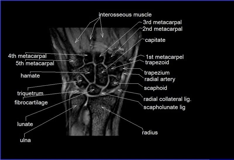
Wrist Mri Anatomy - Radiography is the primary modality used to diagnose and stage arthritis of the hands and wrist.
Wrist Mri Anatomy - 40 public playlists include this case. The complexity of this three arc system creates multiple synergistic joints that allow the wrist a dynamic range of motion. This article aims to frame a general concept of an mri protocol for the assessment of the wrist. Axial views are especially good to visualize tendons, blood vessels, nerves and the two passageways of the radiocarpal joint (carpal tunnel, ulnar canal). Magnetic resonance imaging is particularly well suited for the medical evaluation of the musculoskeletal (msk) system including the knee, shoulder, ankle, wrist and elbow.
A systematic review in the mri of the wrist is essential since wrist anatomy itself is a complex entity with small structures, pathologies and injury patterns that are manifold and involve a whole lot of different therapeutical approaches. Magnetic resonance imaging is particularly well suited for the medical evaluation of the musculoskeletal (msk) system including the knee, shoulder, ankle, wrist and elbow. 2 articles feature images from this case. ( a) schematic diagram shows triangular fibrocartilage complex (tfcc), scapholunate (sl), and lunotriquetral (lt) ligaments. Axial figure 7.1.1 figure 7.1.2 figure 7.1.3 figure 7.1.4 figure 7.1.5 figure 7.1.6 figure 7.1.7 figure 7.1.8 figure 7.1.9 figure 7.1.10 figure 7.1.11 figure 7.1.12. The osseous anatomy of the wrist includes the distal radius and ulna, eight carpal bones, and the five metacarpals (figure 1). The complexity of this three arc system creates multiple synergistic joints that allow the wrist a dynamic range of motion.
Wrist Radiographic Anatomy wikiRadiography
Special attention is paid to key wrist ligaments that play a. ( a) schematic diagram shows triangular fibrocartilage complex (tfcc), scapholunate (sl), and lunotriquetral (lt) ligaments. Magnetic resonance imaging is particularly well suited for the medical evaluation of the musculoskeletal (msk) system including the knee, shoulder, ankle, wrist and elbow. Variant anatomy, imaging pearls, and.
Wrist on 3T MR and 3D pictures normal anatomy eAnatomy
Variant anatomy, imaging pearls, and clinical significance are also discussed. In this manuscript we describe the normal anatomy, imaging techniques, and mri findings of various traumatic and pathologic conditions of the wrist and hand including occult fractures, osteonecrosis, ligamentous and tendon injuries, and entrapment neuropathies. ( b) coronal proton density with fat saturation mr image.
MRI Read wrist joint axial viewcross sectional Anatomy of wrist joint
For a proper radiological interpretation, wrist mri images must be obtained in all three planes; 1) explanation of the optimal coil, sequences and patient positioning 2) demonstration of normal mr anatomy of the wrist, presented by anatomic group (a) osseous morphology and alignment (b) intrinsic ligaments of the wrist (c) triangular fibrocartilage complex (d) extensor.
Anatomy and imaging of wrist joint (MRI AND XRAY)
In this manuscript we describe the normal anatomy, imaging techniques, and mri findings of various traumatic and pathologic conditions of the wrist and hand including occult fractures, osteonecrosis, ligamentous and tendon injuries, and entrapment neuropathies. This article reviews the normal anatomy of the extensor tendons of the wrist as well as the clinical presentation and.
Wrist Mri Anatomy
41 public playlists include this case. Treatment options are also discussed. The osseous anatomy of the wrist includes the distal radius and ulna, eight carpal bones, and the five metacarpals (figure 1). This mri wrist axial cross sectional anatomy tool is absolutely free to use. Axial views are especially good to visualize tendons, blood vessels,.
Wrist Joint Anatomy Concise Medical Knowledge
Normal mri study of the wrist with pd and pd fs sequences in 3 planes. 1) explanation of the optimal coil, sequences and patient positioning 2) demonstration of normal mr anatomy of the wrist, presented by anatomic group (a) osseous morphology and alignment (b) intrinsic ligaments of the wrist (c) triangular fibrocartilage complex (d) extensor.
MRI Wrist Coronal Anatomy Wrist tendon and ligaments Cross sectional
This mri wrist axial cross sectional anatomy tool is absolutely free to use. Axial views are especially good to visualize tendons, blood vessels, nerves and the two passageways of the radiocarpal joint (carpal tunnel, ulnar canal). Mri of the wrist is often challenging because the components of the wrist are small and have complex anatomy..
Wrist Imaging Article
Mri of the wrist includes assessing the wrist’s bony structures, the captured distal radius and ulna to the bases, and proximal parts of the metacarpals (long bones within the hand), including the proximal and distal row of the wrist (carpal) bones (8). This mri wrist axial cross sectional anatomy tool is absolutely free to use..
MRI Wrist Case Study Greater Waterbury Imaging Center
Magnetic resonance imaging is particularly well suited for the medical evaluation of the musculoskeletal (msk) system including the knee, shoulder, ankle, wrist and elbow. Normal radiographic anatomy of the wrist. The mri wrist protocol encompasses a set of mri sequences for the routine assessment of the wrist joint. 2 articles feature images from this case..
6 The Wrist and Hand Diagnostic Imaging Musculoskeletal Key
This mri wrist axial cross sectional anatomy tool is absolutely free to use. Freitasrad is for educational purposes only and should not be used for medical treatment. However, with recent technical advances in extremity coil design, mr imaging has provided us with new insights into the difficult anatomy of the wrist by allowing improved visualization.
Wrist Mri Anatomy ( a ) schematic diagram shows triangular fibrocartilage complex (tfcc), scapholunate (sl), and lunotriquetral (lt) ligaments. 41 public playlists include this case. The mri wrist protocol encompasses a set of mri sequences for the routine assessment of the wrist joint. 40 public playlists include this case. Pinpoints detailed views across anatomical regions & modalities (ct, mri, radiographs), anatomic diagrams and nuclear images.
1) Explanation Of The Optimal Coil, Sequences And Patient Positioning 2) Demonstration Of Normal Mr Anatomy Of The Wrist, Presented By Anatomic Group (A) Osseous Morphology And Alignment (B) Intrinsic Ligaments Of The Wrist (C) Triangular Fibrocartilage Complex (D) Extensor Compartments (E) Carpal Tunnel And Guyon’s Canal 3) Examples Of Commonly.
This article aims to frame a general concept of an mri protocol for the assessment of the wrist. Somewhat confusingly, the term carpus can be used as a synonym for the wrist joint as a whole, or in a more restricted sense to refer to the eight bones of the wrist ( cf. It is the most complete reference of human anatomy available on the web, ios and android devices. Use the mouse scroll wheel to move the images up and down, or alternatively, use the tiny arrows (→) on both sides of the image to navigate through the images.
40 Public Playlists Include This Case.
The complexity of this three arc system creates multiple synergistic joints that allow the wrist a dynamic range of motion. Pinpoints detailed views across anatomical regions & modalities (ct, mri, radiographs), anatomic diagrams and nuclear images. 41 public playlists include this case. This mri wrist axial cross sectional anatomy tool is absolutely free to use.
Treatment Options Are Also Discussed.
5 articles feature images from this case. The wrist is a complex synovial joint formed by articulations of the radius, the articular disc of the distal radioulnar joint and the carpal bones. Mri of the wrist includes assessing the wrist’s bony structures, the captured distal radius and ulna to the bases, and proximal parts of the metacarpals (long bones within the hand), including the proximal and distal row of the wrist (carpal) bones (8). In this manuscript we describe the normal anatomy, imaging techniques, and mri findings of various traumatic and pathologic conditions of the wrist and hand including occult fractures, osteonecrosis, ligamentous and tendon injuries, and entrapment neuropathies.
( B ) Coronal Proton Density With Fat Saturation Mr Image Of The Wrist Shows Tfcc (Long Arrow), Lt Ligament (Short Arrow), And Sl Ligament (Circle).
( a) schematic diagram shows triangular fibrocartilage complex (tfcc), scapholunate (sl), and lunotriquetral (lt) ligaments. Magnetic resonance imaging is particularly well suited for the medical evaluation of the musculoskeletal (msk) system including the knee, shoulder, ankle, wrist and elbow. A systematic review in the mri of the wrist is essential since wrist anatomy itself is a complex entity with small structures, pathologies and injury patterns that are manifold and involve a whole lot of different therapeutical approaches. Protocol specifics will vary depending on mri scanner type, specific hardware and software, radiologist and perhaps referrer preference.


