
Pancreas Ultrasound Anatomy - The pancreas is easily investigated in children thanks to the relative lack of fat tissue and the large left hepatic lobe with an optimal acoustic window.
Pancreas Ultrasound Anatomy - As ultrasound (us) is simple and less invasive than other imaging modalities, this technique is widely used for mass screening. The patient is scanned in a supine position. The errors and mistakes in question were divided into three categories: At least three gray scale images of the right. The pancreas is easily investigated in children thanks to the relative lack of fat tissue and the large left hepatic lobe with an optimal acoustic window.
The anatomy of the pancreas and surrounding structures is critical for understanding the surgical indications and contraindications for pancreatic cancer. Use the splenic vein to help identify the pancreas superficial to this. The pancreas is an extended, accessory digestive gland that is found retroperitoneally, crossing the bodies of the l1 and l2 vertebrae on the posterior abdominal wall. Learn how to use ultrasound to evaluate the pancreas, including pancreas anatomy and physiology and sonographic anatomy of the pancreas. 2) mistakes related to anatomical structures localized in the vicinity of the pancreas (caudate lobe of the liver, other organ. In this way, the presence of bowel gas is limited, the stomach is empty of food, and the entire organ can be visualized. The pancreas is easily investigated in children thanks to the relative lack of fat tissue and the large left hepatic lobe with an optimal acoustic window.

Abdominal ultrasound
The pancreas is a large gland in the back of your abdomen (belly). To put it in a clinical context, its oblique position makes it impossible to see the entire pancreas in a single transverse section. However, visualizing the entire pancreas due to complicated anatomy, obesity and overlying gas can be difficult. Citation, doi, disclosures.
Ultrasound of the Pancreas Radiology Key
It’s part of your digestive system and your endocrine system. Anatomy, physiology & ultrasound appearance :: The pancreas is a large gland in the back of your abdomen (belly). The use of high frequency, even linear transducers, usually results in detailed images of all pancreatic areas. Your pancreas is a dual organ — like a.

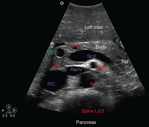
Pancreas Ultrasound Labeled
Often, even with a valiant. It is important to start the ultrasound examination with an evaluation of the pancreas before the child swallows air by talking or crying, and before air normally migrates toward the gastric antrum and transverse colon. However, since its echogenicity varies with age and disease process, it is most useful to.

Pancreas Ultrasound Labeled
The patient is scanned in a supine position. Often, even with a valiant. To put it in a clinical context, its oblique position makes it impossible to see the entire pancreas in a single transverse section. Thus the spleen can be used as a window and a left intercostal coronal approach can. The use of.

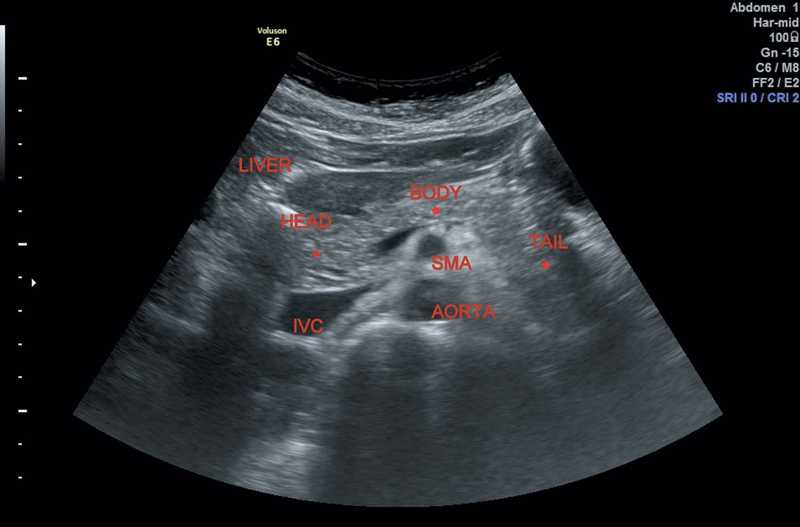
Normal Pancreas Ultrasound
Strategies to see the pancreas include using the left hepatic lobe as an acoustic window, imaging an npo patient (not usually possible), or pressing on the abdomen firmly to push away overlying air (not always comfortable). 2) mistakes related to anatomical structures localized in the vicinity of the pancreas (caudate lobe of the liver, other.

Ultrasound of pancrease in Radiology
In this way, the presence of bowel gas is limited, the stomach is empty of food, and the entire organ can be visualized. Anatomy should be intentionally imaged in an organized fashion and correctly labeled for clarity. It is important to start the ultrasound examination with an evaluation of the pancreas before the child swallows.
Pancreas Radiology Key
However, since its echogenicity varies with age and disease process, it is most useful to rely on the adjacent anatomic structures as landmarks when tracing the gland in the transverse and longitudinal orientations. In this way, the presence of bowel gas is limited, the stomach is empty of food, and the entire organ can be.
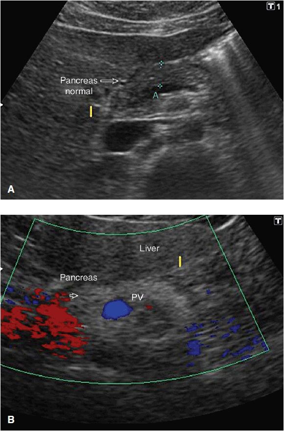
Ultrasound of the Pancreas Radiology Key
Download reference work entry pdf. Your pancreas is a dual organ — like a factory with two production lines. The longest part of the pancreas is located to the left of the midline, with the tail near the splenic hilum usually slightly above the head. 1) mistakes related to the anatomical structure of the pancreas.

Pancreas Ultrasound Anatomy
The pancreas can be visualized at us in most patients, independent of gastrointestinal gas interference and fat. In this way, the presence of bowel gas is limited, the stomach is empty of food, and the entire organ can be visualized. As ultrasound (us) is simple and less invasive than other imaging modalities, this technique is.

Pancreas Ultrasound Labeled
The errors and mistakes in question were divided into three categories: To put it in a clinical context, its oblique position makes it impossible to see the entire pancreas in a single transverse section. The patient is scanned in a supine position. It’s part of your digestive system and your endocrine system. 12k views 2.
Pancreas Ultrasound Anatomy Thus the spleen can be used as a window and a left intercostal coronal approach can. The patient is scanned in a supine position. Pancreas anatomy & physiology ultrasound training. The pancreas is an unpaired accessory digestive gland. The pancreas is distinguished on ultrasound by its uniquely glandular parenchyma;
Cystic Pancreatic Lesions Are Increasingly Identified Due To.
The anatomy of the pancreas and surrounding structures is critical for understanding the surgical indications and contraindications for pancreatic cancer. Your pancreas is a dual organ — like a factory with two production lines. Ultrasound is the first modality used in the evaluation of pancreatic disease. Use the splenic vein to help identify the pancreas superficial to this.
The Pancreas Lies Transversely In The Upper Abdomen Between The Duodenum On The Right And The Spleen On The Left.
It’s part of your digestive system and your endocrine system. The pancreas can be visualized at us in most patients, independent of gastrointestinal gas interference and fat. Strategies to see the pancreas include using the left hepatic lobe as an acoustic window, imaging an npo patient (not usually possible), or pressing on the abdomen firmly to push away overlying air (not always comfortable). It is important to start the ultrasound examination with an evaluation of the pancreas before the child swallows air by talking or crying, and before air normally migrates toward the gastric antrum and transverse colon.
The Patient Is Scanned In A Supine Position.
The pancreas is an unpaired accessory digestive gland. The longest part of the pancreas is located to the left of the midline, with the tail near the splenic hilum usually slightly above the head. At least three gray scale images of the left lobe to include lateral, mid, medial. To put it in a clinical context, its oblique position makes it impossible to see the entire pancreas in a single transverse section.
The Use Of High Frequency, Even Linear Transducers, Usually Results In Detailed Images Of All Pancreatic Areas.
Anatomy, physiology & ultrasound appearance :: 2) mistakes related to anatomical structures localized in the vicinity of the pancreas (caudate lobe of the liver, other organ. Learn how to use ultrasound to evaluate the pancreas, including pancreas anatomy and physiology and sonographic anatomy of the pancreas. 12k views 2 years ago normal abdomen.