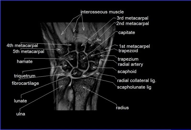
Mri Anatomy Wrist - It is the most complete reference of human anatomy available on the web, ios and android.
Mri Anatomy Wrist - Web this article aims to review the normal and pathologic appearance of intrinsic and extrinsic wrist ligaments with a focus on mri. Anatomy arthrogram anatomy basic shoulder mri. 2 articles feature images from this case. Web the wrist is the part of the upper limb between the forearm and the palm. Web in this manuscript we describe the normal anatomy, imaging techniques, and mri findings of various traumatic and pathologic conditions of the wrist and hand.
Web the complex anatomy of the wrist can be demonstrated by mri. Web the typical mri appearance consists of t1 intermediate and t2 hyperintense foci, which demonstrate mild enhancement with a peripheral and septal pattern after administration. Web the mri wrist protocol encompasses a set of mri sequences for the routine assessment of the wrist joint. 41 public playlists include this case. It is the most complete reference of human anatomy available on the web, ios and android. Web this mri wrist axial cross sectional anatomy tool is absolutely free to use. Normal mri study of the wrist with pd and pd fs sequences in 3 planes.
Mri Anatomy Of Wrist
( a) schematic diagram shows triangular fibrocartilage complex (tfcc), scapholunate (sl), and lunotriquetral (lt) ligaments. Web the mri wrist protocol encompasses a set of mri sequences for the routine assessment of the wrist joint. Web anatomy basic knee mri checklist. Web axial figure 7.1.1 figure 7.1.2 figure 7.1.3 figure 7.1.4 figure 7.1.5 figure 7.1.6 figure.
Wrist/Hand MRI
Normal mri study of the wrist with pd and pd fs sequences in 3 planes. This article aims to frame a general concept of an. Web axial figure 7.1.1 figure 7.1.2 figure 7.1.3 figure 7.1.4 figure 7.1.5 figure 7.1.6 figure 7.1.7 figure 7.1.8 figure 7.1.9 figure 7.1.10 figure 7.1.11 figure 7.1.12. Web this mri wrist.
Wrist on 3T MR and 3D pictures normal anatomy eAnatomy
Web the typical mri appearance consists of t1 intermediate and t2 hyperintense foci, which demonstrate mild enhancement with a peripheral and septal pattern after administration. Web the mri wrist protocol encompasses a set of mri sequences for the routine assessment of the wrist joint. Magnetic resonance imaging is particularly well suited for the medical evaluation.
MRI Wrist Coronal Anatomy Wrist tendon and ligaments Cross sectional
( a) schematic diagram shows triangular fibrocartilage complex (tfcc), scapholunate (sl), and lunotriquetral (lt) ligaments. Osteoarthritis may incidentally be seen on mri as areas of. Web this article aims to review the normal and pathologic appearance of intrinsic and extrinsic wrist ligaments with a focus on mri. Web the wrist is the part of the.
Wrist Mri Anatomy
Web the typical mri appearance consists of t1 intermediate and t2 hyperintense foci, which demonstrate mild enhancement with a peripheral and septal pattern after administration. Web anatomy basic knee mri checklist. Use the mouse scroll wheel to move the images up and down, or alternatively, use the tiny arrows (→). Web wrist/hand anatomy & scanning.
Wrist Radiographic Anatomy wikiRadiography
This article aims to frame a general concept of an. Web the mri wrist protocol encompasses a set of mri sequences for the routine assessment of the wrist joint. Web anatomy basic knee mri checklist. ( a) schematic diagram shows triangular fibrocartilage complex (tfcc), scapholunate (sl), and lunotriquetral (lt) ligaments. Web the typical mri appearance.
Anatomy and imaging of wrist joint (MRI AND XRAY)
Web the wrist is a complex synovial joint formed by articulations of the radius, the articular disc of the distal radioulnar joint and the carpal bones. Web the mri wrist protocol encompasses a set of mri sequences for the routine assessment of the wrist joint. The bony framework consists of a wrist joint (radiocarpal joint)..
MRI Wrist Case Study Greater Waterbury Imaging Center
Web the wrist is the part of the upper limb between the forearm and the palm. Web in this manuscript we describe the normal anatomy, imaging techniques, and mri findings of various traumatic and pathologic conditions of the wrist and hand. Magnetic resonance imaging is particularly well suited for the medical evaluation of the musculoskeletal.
MRI Read wrist joint axial viewcross sectional Anatomy of wrist joint
Osteoarthritis may incidentally be seen on mri as areas of. ( a) schematic diagram shows triangular fibrocartilage complex (tfcc), scapholunate (sl), and lunotriquetral (lt) ligaments. 2 articles feature images from this case. The bony framework consists of a wrist joint (radiocarpal joint). Web the complex anatomy of the wrist can be demonstrated by mri. Magnetic.
Wrist Anatomy X Ray Labeled
Web wrist/hand anatomy & scanning techniques. ( a) schematic diagram shows triangular fibrocartilage complex (tfcc), scapholunate (sl), and lunotriquetral (lt) ligaments. Web anatomy basic knee mri checklist. 2 articles feature images from this case. Web radiography is the primary modality used to diagnose and stage arthritis of the hands and wrist. Web the complex anatomy.
Mri Anatomy Wrist Use the mouse scroll wheel to move the images up and down, or alternatively, use the tiny arrows (→). Web on this module on the anatomy of the wrist in 3t mri, we labeled over 340 anatomical structures in 208 mr images of the wrist and 3d reconstructions of the. Web this approach is an example of how to create a radiological report of an mri of the wrist with coverage of the most common anatomical sites of possible pathology,. Web the typical mri appearance consists of t1 intermediate and t2 hyperintense foci, which demonstrate mild enhancement with a peripheral and septal pattern after administration. Web this mri wrist axial cross sectional anatomy tool is absolutely free to use.
Magnetic Resonance Imaging Is Particularly Well Suited For The Medical Evaluation Of The Musculoskeletal (Msk) System Including The Knee, Shoulder, Ankle,.
Web the mri wrist protocol encompasses a set of mri sequences for the routine assessment of the wrist joint. Web axial figure 7.1.1 figure 7.1.2 figure 7.1.3 figure 7.1.4 figure 7.1.5 figure 7.1.6 figure 7.1.7 figure 7.1.8 figure 7.1.9 figure 7.1.10 figure 7.1.11 figure 7.1.12. Osteoarthritis may incidentally be seen on mri as areas of. Web the typical mri appearance consists of t1 intermediate and t2 hyperintense foci, which demonstrate mild enhancement with a peripheral and septal pattern after administration.
Normal Mri Study Of The Wrist With Pd And Pd Fs Sequences In 3 Planes.
Web on this module on the anatomy of the wrist in 3t mri, we labeled over 340 anatomical structures in 208 mr images of the wrist and 3d reconstructions of the. Web wrist/hand anatomy & scanning techniques. Web anatomy basic knee mri checklist. Use the mouse scroll wheel to move the images up and down, or alternatively, use the tiny arrows (→).
Web The Wrist Is A Complex Synovial Joint Formed By Articulations Of The Radius, The Articular Disc Of The Distal Radioulnar Joint And The Carpal Bones.
Web the wrist is the part of the upper limb between the forearm and the palm. The bony framework consists of a wrist joint (radiocarpal joint). 2 articles feature images from this case. It is the most complete reference of human anatomy available on the web, ios and android.
Web Mri Of The Wrist Is Commonly Performed To Evaluate Individuals With Wrist Pain, Allowing Detailed Anatomic Evaluation And Accurate Characterization Of Wrist.
Anatomy arthrogram anatomy basic shoulder mri. Web this approach is an example of how to create a radiological report of an mri of the wrist with coverage of the most common anatomical sites of possible pathology,. Web in this manuscript we describe the normal anatomy, imaging techniques, and mri findings of various traumatic and pathologic conditions of the wrist and hand. 41 public playlists include this case.


