
Lateral Cxr Anatomy - Web the white “3” lies on the posterior 3rd rib while the black “3” lies on the anterior 3 rd rib.
Lateral Cxr Anatomy - The right hemidiaphragm is slightly higher than the left. This article outlines the key features of the lateral view, such as the hilum, the airway, and the three darkenings, and explains their normal and abnormal appearances. The stomach and spleen are located inferior to the left hemidiaphragm. Why are two views of the chest done? If the side is not specified, a left lateral is usually taken.
Two views (pa and lateral) are obtained at 90 degrees from each other in order to gain an appreciation of dimensionality. Ra = right atrium, rv = right ventricle, la = left atrium, lv = left ventricle, vcs = vena cava superior or superior caval vein. Provide a systemic approach to reviewing the lateral cxr. Highlight essential review areas which often exhibit pathology that is Web the lateral chest radiograph is an imaging study that can be challenging to both the novice or seasoned radiologist. The stomach and spleen are located inferior to the left hemidiaphragm. Web the lungs and pleural spaces are clear.

Lateral Cxr Anatomy
The liver is located inferior to the right hemidiaphragm. This can be helpful in settings where the single view is limited in localizing pathology (i.e. Identifying the exact lobe of a lobar pneumonia in the right lung). This is usually done together with a view from the front of the chest, also called a frontal.

Normal Lateral Chest X Ray Male Online Image
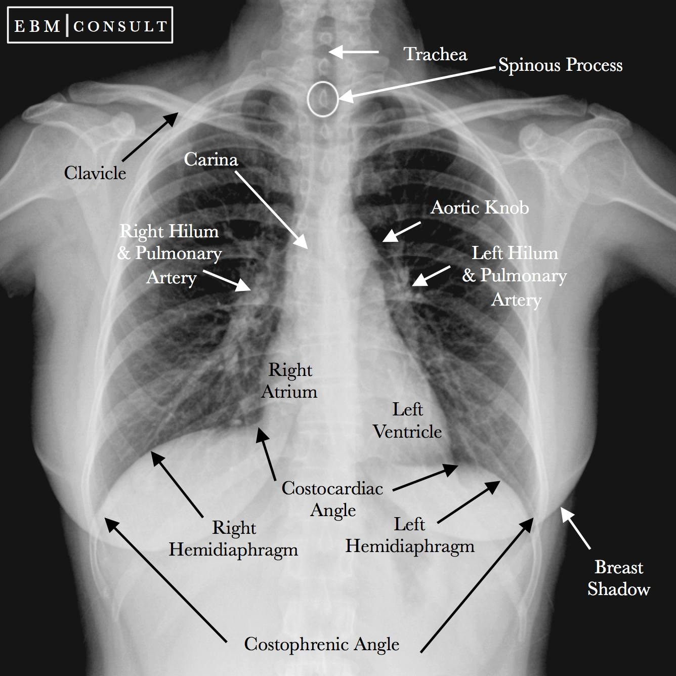
Anatomy of lateral chest radiograph is shown below: This article outlines the key features of the lateral view, such as the hilum, the airway, and the three darkenings, and explains their normal and abnormal appearances. Provide a systemic approach to reviewing the lateral cxr. This tutorial covers the trachea, lungs, heart, diaphragm, ribs, scapulae, breasts,.

Lateral Chest X Ray Anatomy Anatomical Charts & Posters
This article outlines the key features of the lateral view, such as the hilum, the airway, and the three darkenings, and explains their normal and abnormal appearances. In this paper we offer a brief guide to demystify and clarify this cheap and useful investigation. These images are read together. Web identify normal anatomical structures on.

Normal Lateral Chest Radiograph
Provide a systemic approach to reviewing the lateral cxr. In this paper we offer a brief guide to demystify and clarify this cheap and useful investigation. The patient is facing towards the left on the lateral view. Why are two views of the chest done? Web identify normal anatomical structures on the lateral cxr. Highlight.

Labelled Normal Chest XRay (CXR) Radiology, Radiology student
Anatomy of lateral chest radiograph is shown below: If the side is not specified, a left lateral is usually taken. Identifying the exact lobe of a lobar pneumonia in the right lung). Why are two views of the chest done? The objective evaluation is based on the relative positioning and size of the lv (white.

Lateral Chest Radiograph Anatomy
This is usually done together with a view from the front of the chest, also called a frontal view. In this paper we offer a brief guide to demystify and clarify this cheap and useful investigation. Anatomy of lateral chest radiograph is shown below: If the side is not specified, a left lateral is usually.

Normal Chest XRay • LITFL Medical Blog • Labelled Radiology
Anatomy of lateral chest radiograph is shown below: Identifying the exact lobe of a lobar pneumonia in the right lung). Web the white “3” lies on the posterior 3rd rib while the black “3” lies on the anterior 3 rd rib. Ra = right atrium, rv = right ventricle, la = left atrium, lv =.
Lateral Chest X Ray Anatomy Anatomical Charts & Posters
Anatomy of lateral chest radiograph is shown below: The pa exam is viewed as if the patient is standing in front of you with their right side on your left. The right hemidiaphragm is slightly higher than the left. Two views (pa and lateral) are obtained at 90 degrees from each other in order to.

Chest Radiographic Anatomy wikiRadiography
These images are read together. Web the white “3” lies on the posterior 3rd rib while the black “3” lies on the anterior 3 rd rib. This tutorial covers the trachea, lungs, heart, diaphragm, ribs, scapulae, breasts, bowel gas and more. Web the lateral chest radiograph is an imaging study that can be challenging to.

Lateral Chest X Ray Anatomy Anatomical Charts & Posters
This can be helpful in settings where the single view is limited in localizing pathology (i.e. The liver is located inferior to the right hemidiaphragm. The pa exam is viewed as if the patient is standing in front of you with their right side on your left. Provide a systemic approach to reviewing the lateral.
Lateral Cxr Anatomy Two views (pa and lateral) are obtained at 90 degrees from each other in order to gain an appreciation of dimensionality. These images are read together. Web learn how to analyze the lateral chest radiograph with a simple and effective method. This tutorial covers the trachea, lungs, heart, diaphragm, ribs, scapulae, breasts, bowel gas and more. The patient is facing towards the left on the lateral view.
Identifying The Exact Lobe Of A Lobar Pneumonia In The Right Lung).
The hemidiaphragms are domed structures. Highlight essential review areas which often exhibit pathology that is The stomach and spleen are located inferior to the left hemidiaphragm. Web the lateral chest radiograph is an imaging study that can be challenging to both the novice or seasoned radiologist.
Learn The Anatomy That Lurks Within The Lateral Chest Radiograph, And Tips On How To Read Them With Aplomb.
Two views (pa and lateral) are obtained at 90 degrees from each other in order to gain an appreciation of dimensionality. This article outlines the key features of the lateral view, such as the hilum, the airway, and the three darkenings, and explains their normal and abnormal appearances. Anatomy of lateral chest radiograph is shown below: This tutorial covers the trachea, lungs, heart, diaphragm, ribs, scapulae, breasts, bowel gas and more.
The Patient Is Facing Towards The Left On The Lateral View.
Why are two views of the chest done? This is usually done together with a view from the front of the chest, also called a frontal view. Web identify normal anatomical structures on the lateral cxr. If the side is not specified, a left lateral is usually taken.
The Liver Is Located Inferior To The Right Hemidiaphragm.
In this paper we offer a brief guide to demystify and clarify this cheap and useful investigation. The pa exam is viewed as if the patient is standing in front of you with their right side on your left. Ra = right atrium, rv = right ventricle, la = left atrium, lv = left ventricle, vcs = vena cava superior or superior caval vein. Web the white “3” lies on the posterior 3rd rib while the black “3” lies on the anterior 3 rd rib.