
Internal Anatomy Of The Perch - Use scissors to make the cuts through skin and muscle shown in figure 1.
Internal Anatomy Of The Perch - Begin the incision along the dorsal side of the fish no higher than the lateral line. To examine the internal and external anatomy of the perch. They are the largest group of vertebrates; Each letter corresponds to an internal body part, a: Label the upper jaw (maxilla) and the.
In this laboratory you will observe the internal and external structures of a perch, a typical bony fish. Cells often become specialized to perform certain functions. This video details the internal anatomy of a perch. For more detailed dissection instructions and. Use dissecting pins to secure the fish to the dissecting pan. A tissue is a group of similar cells performing a similar function (fig. The flesh of this fish is highly valued.

Internal Perch Anatomy Anatomical Charts & Posters
1) stabilize against rolling or sudden turns. Most of the organs reside in the ventral half of the fish’s body. Auricle of the heart, c: Use dissecting pins to secure the fish to the dissecting pan. Observing the fish’s internal anatomy. Phylum chordata, subphylum vertebrata, c. They are the largest group of vertebrates; Ventricle of.

perch dissection2
This video details the internal anatomy of a perch. Yellow perch are primarily bottom feeders with a slow deliberate bite. Learn vocabulary, terms, and more with flashcards, games, and other study tools. 7) holds exoskeleton and nerve system. A tissue is a group of similar cells performing a similar function (fig. Below is a brief.
Internal Perch Anatomy Anatomical Charts & Posters
Most of the organs reside in the ventral half of the fish’s body. Each individual system can be viewed and discussed separately. 7) holds exoskeleton and nerve system. This is intended for college level biologists. In this laboratory you will observe the internal and external structures of a perch, a typical bony fish. Auricle of.

PPT Perch Dissection PreLab PowerPoint Presentation ID3111478
Label the upper jaw (maxilla) and the. Use dissecting pins to secure the fish to the dissecting pan. Start studying internal anatomy of perch. Begin the incision along the dorsal side of the fish no higher than the lateral line. Studies the external and internal anatomy of a perch, a bony fish, and compares male.

Perch Internal Anatomy
Secure the fish to the dissecting pan. Perch are a great way to learn about bony. Each letter corresponds to an internal body part, a: For example, muscle cells contract, nerve cells transmit impulses, and gland cells produce chemicals. After making the cuts, carefully lift off the flap of skin and muscle to expose the.

internal stuff perch part 1 Diagram Quizlet
Ventricle of the heart, d: Internal fish anatomy and the function of fish organ systems. Use dissecting pins to secure the fish to the dissecting pan. Use scissors to make the cuts through skin and muscle shown in figure 1. 7) holds exoskeleton and nerve system. This program facilitates a study of the anatomy of.

perch anatomy (internal) Diagram Quizlet
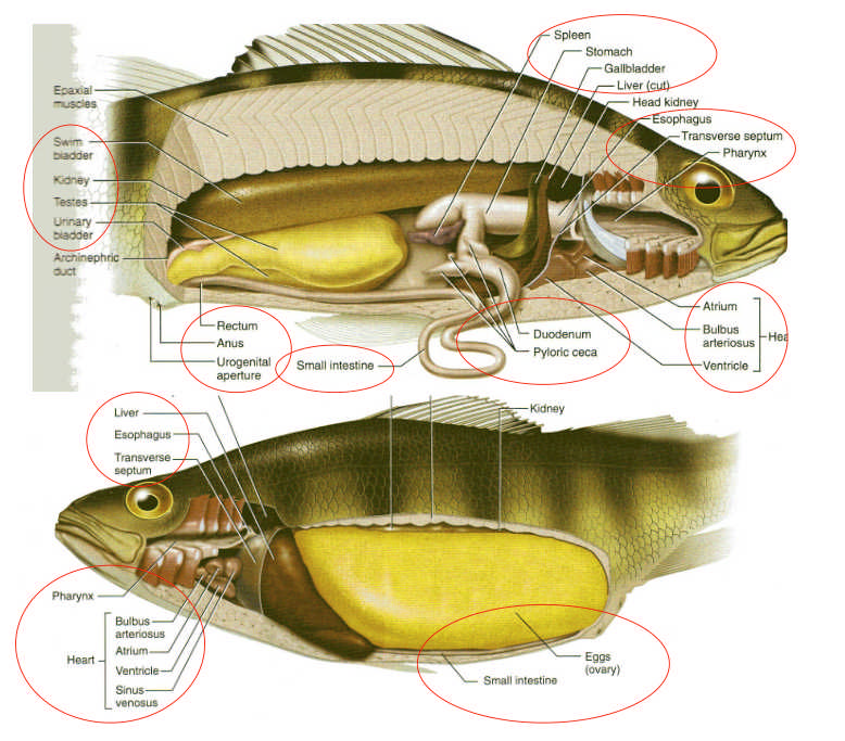
Use dissecting pins to secure the fish to the dissecting pan. Anatomy of a perch perch: Internal anatomy of a perch using a model. Perch are a great way to learn about bony. The above picture is a labeled image of the internal anatomy of the species perch perca flavescens. For example, muscle cells contract,.

PPT Perch Dissection PreLab PowerPoint Presentation, free download
After making the cuts, carefully lift off the flap of skin and muscle to expose the internal organs in the body cavity. In this laboratory you will observe the internal and external structures of a perch, a typical bony fish. Use scissors to make the cuts through skin and muscle shown in figure 4. This.

Perch Diagram 3 Diagram Quizlet
Living things are composed of cells. You will see how its structures make it ideally suited to living in a aquatic environment. Study with quizlet and memorize flashcards containing terms. Start studying internal anatomy of perch. Auricle of the heart, c: Internal fish anatomy and the function of fish organ systems. Cells often become specialized.

Perch Dissection
Figure 3 shows the incisions to be made for viewing the internal structures of the perch. Yellow perch are primarily bottom feeders with a slow deliberate bite. 5) aids in going up or down. Learn how to dissect a perch in this video, which also covers its external and internal anatomy and physiology. Each letter.
Internal Anatomy Of The Perch 7) holds exoskeleton and nerve system. Use scissors to make the cuts through skin and muscle shown in figure 4. Below is a brief survey of the internal and external anatomy of the perch. To examine the internal and external anatomy of the perch. Ventricle of the heart, d:
Label The Upper Jaw (Maxilla) And The.
Use scissors to make the cuts through skin and muscle shown in figure 4. Internal anatomy of a perch using a model. 5) aids in going up or down. Learn vocabulary, terms, and more with flashcards, games, and other study tools.
This Video Details The Internal Anatomy Of A Perch.
Anatomy of a perch perch: 4) maintains depth or flight or walking. Ventricle of the heart, d: Each individual system can be viewed and discussed separately.
Auricle Of The Heart, C:
Use dissecting pins to secure the fish to the dissecting pan. Start studying internal anatomy of perch. Perch are vertebrates in a group called the “ray finned fishes” because they have rays/spines in their fins. Label the anterior, posterior, dorsal, and ventral sides on figure 1.
They Eat Almost Anything, But Prefer Minnows, Insect Larvae, Plankton, And Worms.
Open the mouth and observe its jaws. They are the largest group of vertebrates; Use scissors to make the cuts through skin and muscle shown in figure 1. The above picture is a labeled image of the internal anatomy of the species perch perca flavescens.