
Horse Distal Limb Anatomy - I find this a welcome addition to an atlas of anatomy.
Horse Distal Limb Anatomy - Learn about the anatomy and imaging of the equine distal limb with interactive 3d models, videos and quizzes from the royal veterinary college. Over time, two toes disappeared completely, whilst. Home 3d radiographic projection select a body part and angle on the left, then select the type of image from the top menu. There is no muscle below the knee and hock. The distal limb is everything below the knee and the hock.
The author uses a variety of diagnostic modalities to illustrate the normal anatomy of the equine distal limb. There is no muscle below the knee and hock. The distal limb is everything below the knee and the hock. I find this a welcome addition to an atlas of anatomy. This interactive anatomy of the equine distal limb is designed to increase the users familiarity with anatomical structures and also to allow examination of all surfaces of bones, all regions of blood supply, and the relations of anatomical structures to the complete foot. Home 3d radiographic projection select a body part and angle on the left, then select the type of image from the top menu. Over time, two toes disappeared completely, whilst.
horse limb anatomy
The distal limb refers to the horse’s lower leg, below the carpus (knee) or hock. The author uses a variety of diagnostic modalities to illustrate the normal anatomy of the equine distal limb. Over time, two toes disappeared completely, whilst. Diagnosis and management of distal limb lameness require a precise knowledge of the functional anatomy.

Understanding Navicular Syndrome & Heel Pain in Horses
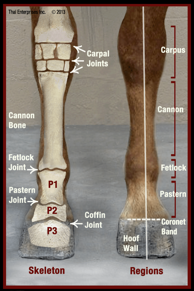
Over time, two toes disappeared completely, whilst. I find this a welcome addition to an atlas of anatomy. The distal limb refers to the horse’s lower leg, below the carpus (knee) or hock. There is no muscle below the knee and hock. There are nine bones total and each plays a vital role in movement.

Equine distal limb anatomy print Plastination Anatomy Embedding
The distal limb refers to the horse’s lower leg, below the carpus (knee) or hock. The distal limb bones are the foundation of equine lower leg. Learn about the anatomy and imaging of the equine distal limb with interactive 3d models, videos and quizzes from the royal veterinary college. Home 3d radiographic projection select a.

Equine Distal Limb Stunning full color anatomy photos that reveal the
Home 3d radiographic projection select a body part and angle on the left, then select the type of image from the top menu. Over time, two toes disappeared completely, whilst. The distal limb bones are the foundation of equine lower leg. The distal limb is everything below the knee and the hock. I find this.

Distal hind limb anatomy Horse health, Horse anatomy, Vet medicine
Diagnosis and management of distal limb lameness require a precise knowledge of the functional anatomy and biomechanics of the equine distal joints, ligaments and tendons, presented in the last chapter. There is no muscle below the knee and hock. I find this a welcome addition to an atlas of anatomy. Home 3d radiographic projection select.

Distal limb anatomy Horse anatomy, Horse care, Horses
The distal limb refers to the horse’s lower leg, below the carpus (knee) or hock. Diagnosis and management of distal limb lameness require a precise knowledge of the functional anatomy and biomechanics of the equine distal joints, ligaments and tendons, presented in the last chapter. The author uses a variety of diagnostic modalities to illustrate.

Equine Distal Limb Anatomical illustration
The author uses a variety of diagnostic modalities to illustrate the normal anatomy of the equine distal limb. Home 3d radiographic projection select a body part and angle on the left, then select the type of image from the top menu. Learn about the anatomy and imaging of the equine distal limb with interactive 3d.

Equine distal limb sectional anatomy hoof Plastination Anatomy Embedding
This interactive anatomy of the equine distal limb is designed to increase the users familiarity with anatomical structures and also to allow examination of all surfaces of bones, all regions of blood supply, and the relations of anatomical structures to the complete foot. I find this a welcome addition to an atlas of anatomy. The.

Tendon and ligament physiology Veterian Key
The distal limb bones are the foundation of equine lower leg. There are nine bones total and each plays a vital role in movement and stability. Learn about the anatomy and imaging of the equine distal limb with interactive 3d models, videos and quizzes from the royal veterinary college. Over time, two toes disappeared completely,.

Horse Distal Limb Anatomy
The distal limb bones are the foundation of equine lower leg. Home 3d radiographic projection select a body part and angle on the left, then select the type of image from the top menu. Over time, two toes disappeared completely, whilst. I find this a welcome addition to an atlas of anatomy. Learn about the.
Horse Distal Limb Anatomy There is no muscle below the knee and hock. Diagnosis and management of distal limb lameness require a precise knowledge of the functional anatomy and biomechanics of the equine distal joints, ligaments and tendons, presented in the last chapter. The distal limb is everything below the knee and the hock. The author uses a variety of diagnostic modalities to illustrate the normal anatomy of the equine distal limb. Learn about the anatomy and imaging of the equine distal limb with interactive 3d models, videos and quizzes from the royal veterinary college.
This Interactive Anatomy Of The Equine Distal Limb Is Designed To Increase The Users Familiarity With Anatomical Structures And Also To Allow Examination Of All Surfaces Of Bones, All Regions Of Blood Supply, And The Relations Of Anatomical Structures To The Complete Foot.
Home 3d radiographic projection select a body part and angle on the left, then select the type of image from the top menu. Diagnosis and management of distal limb lameness require a precise knowledge of the functional anatomy and biomechanics of the equine distal joints, ligaments and tendons, presented in the last chapter. The distal limb refers to the horse’s lower leg, below the carpus (knee) or hock. Over time, two toes disappeared completely, whilst.
Learn About The Anatomy And Imaging Of The Equine Distal Limb With Interactive 3D Models, Videos And Quizzes From The Royal Veterinary College.
The distal limb is everything below the knee and the hock. I find this a welcome addition to an atlas of anatomy. The author uses a variety of diagnostic modalities to illustrate the normal anatomy of the equine distal limb. There are nine bones total and each plays a vital role in movement and stability.
The Distal Limb Bones Are The Foundation Of Equine Lower Leg.
There is no muscle below the knee and hock.