
Chest X Ray Anatomy Lateral - Figures 3.1 and 3.2 are example pa and lateral views without pathology.
Chest X Ray Anatomy Lateral - The interpretation of a chest film requires the understanding of basic principles. Click image to see overlay from central to peripheral the airway consists of: A lateral view of the chest can show more abnormalities like pneumonia, nodules and masses compared with just a frontal view of the chest. In fact every radiologst should be an expert in chest film reading. The left lung has two lobes and the right has three.
Some of the key topics are pneumonia, atelectasis, congestive heart failure and pneumothorax. Figures 3.1 and 3.2 are example pa and lateral views without pathology. The oblique fissures may be seen on a normal lateral view. The lateral chest radiograph is particularly useful for the recognition of specific chamber enlargement (other than that of the right atrium) for the recognition of the valve prostheses and rings as they are projected away from the spine. Cardiovascular anatomy of the mediastinum on a lateral chest radiograph. In fact every radiologst should be an expert in chest film reading. Imagine you are holding a flashlight, pointing it so that the circle of light appears against a white wall a foot away from you.

Lateral Chest X Ray Anatomy Anatomical Charts & Posters
The pa exam is viewed as if the patient is standing in front of you with their right side on your left. The pa (posterior to anterior) and the lateral. This is generally relevant only in interpretation of a posteroanterior (pa) or anteroposterior (ap) projection and not of the lateral projection. The horizontal fissure (right).

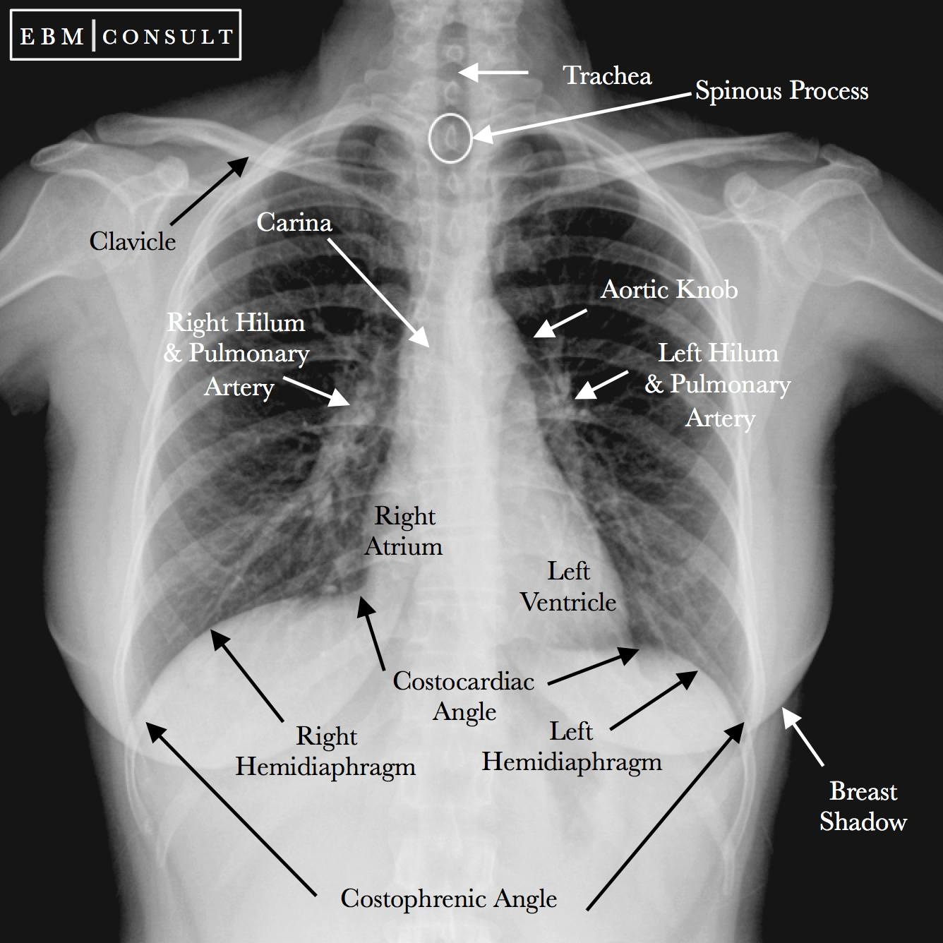
Normal Chest XRay • LITFL Medical Blog • Labelled Radiology
The lateral chest radiograph is a valuable source of information that has become increasingly undervalued in the era of chest computed tomography. What does the lateral view of the chest show? The interpretation of a chest film requires the understanding of basic principles. Each hemidiaphragm should appear as a smooth, domed contour. Imagine you are.

Normal Lateral Chest Radiograph
The lateral chest radiograph is particularly useful for the recognition of specific chamber enlargement (other than that of the right atrium) for the recognition of the valve prostheses and rings as they are projected away from the spine. This orthogonal view to a frontal chest radiograph may be performed as an adjunct in cases where.

Lateral Chest X Ray Anatomy Anatomical Charts & Posters
First is an overview, followed by analysis of the airway and major hilar structures. These images are read together. The diaphragm separates the lungs from the abdominal organs. Each lobe has its own pleural covering. The pa exam is viewed as if the patient is standing in front of you with their right side on.

Lateral Chest X Ray Anatomy Anatomical Charts & Posters
Click image to see overlay from central to peripheral the airway consists of: Other figures in this chapter will show you the different elements of the normal anatomy as they appear in a normal cxr. Sometimes pneumonia can hide behind the heart or. Rotation of a chest radiograph can simulate common pathological processes and make.
Lateral Chest X Ray Anatomy Anatomical Charts & Posters
Trachea, main bronchus, bronchioli and alveoli. The right hemidiaphragm is usually a little higher than the left. The left lung has two lobes and the right has three. First is an overview, followed by analysis of the airway and major hilar structures. Click image to see overlay from central to peripheral the airway consists of:.
Lateral Chest X Ray Anatomy Anatomical Charts & Posters
The lateral chest radiograph is particularly useful for the recognition of specific chamber enlargement (other than that of the right atrium) for the recognition of the valve prostheses and rings as they are projected away from the spine. Sometimes pneumonia can hide behind the heart or. Let’s take a second to try to understand why.

Chest Radiographic Anatomy wikiRadiography
Learn the anatomy that lurks within the lateral chest radiograph, and tips on how to read them with aplomb. The right hemidiaphragm is usually a little higher than the left. Optimal use of the lateral radiograph requires systematic analysis. The diaphragm separates the lungs from the abdominal organs. The lateral chest view can be particularly.

Xrays Concise Medical Knowledge
Each hemidiaphragm should appear as a smooth, domed contour. Each lobe has its own pleural covering. Click image to see overlay from central to peripheral the airway consists of: They are often known as a portable film when performed with a mobile unit. First is an overview, followed by analysis of the airway and major.

Lateral radiograph of the chest The BMJ
Trachea, main bronchus, bronchioli and alveoli. The lateral chest view can be particularly useful in assessing the retrosternal and retrocardiac airspaces. The lateral chest radiograph is particularly useful for the recognition of specific chamber enlargement (other than that of the right atrium) for the recognition of the valve prostheses and rings as they are projected.
Chest X Ray Anatomy Lateral The lateral chest view examines the lungs, bony thoracic cavity, mediastinum, and great vessels. The horizontal fissure (right) is often seen on a normal frontal view. The lateral chest radiograph is a valuable source of information that has become increasingly undervalued in the era of chest computed tomography. Let’s take a second to try to understand why it is that the heart appears bigger than normal on ap studies. They are often known as a portable film when performed with a mobile unit.
Click Image To See Overlay From Central To Peripheral The Airway Consists Of:
In this paper we offer a brief guide to demystify and clarify this cheap and useful investigation. First is an overview, followed by analysis of the airway and major hilar structures. The oblique fissures may be seen on a normal lateral view. A lateral view of the chest can show more abnormalities like pneumonia, nodules and masses compared with just a frontal view of the chest.
The Diaphragm Separates The Lungs From The Abdominal Organs.
This orthogonal view to a frontal chest radiograph may be performed as an adjunct in cases where there is diagnostic uncertainty. The lateral chest view can be particularly useful in assessing the retrosternal and retrocardiac airspaces. The lateral view of the chest is usually performed erect left lateral and labeled with the side closest to the cassette. What does the lateral view of the chest show?
Learn The Anatomy That Lurks Within The Lateral Chest Radiograph, And Tips On How To Read Them With Aplomb.
Trachea, main bronchus, bronchioli and alveoli. The right hemidiaphragm is usually a little higher than the left. The pa exam is viewed as if the patient is standing in front of you with their right side on your left. The lateral chest radiograph is particularly useful for the recognition of specific chamber enlargement (other than that of the right atrium) for the recognition of the valve prostheses and rings as they are projected away from the spine.
Anteroposterior (Ap) Chest Radiographs Can Be Made In The Intensive Care Unit, The Operating Suite, Or The Patient’s Room Using Mobile Equipment.
Each lobe has its own pleural covering. Other figures in this chapter will show you the different elements of the normal anatomy as they appear in a normal cxr. Cardiovascular anatomy of the mediastinum on a lateral chest radiograph. Let’s take a second to try to understand why it is that the heart appears bigger than normal on ap studies.