
Canine Hip Anatomy - Features of the anatomical model include normal right coxofemoral joint, left coxofemoral joint showing hip dysplasia and osteoarthritis, disc with herniation and spinal cord with nerves.
Canine Hip Anatomy - Organs and structures of different regions of a dog. Learn about the veterinary topic of components of the musculoskeletal system in dogs. The canine hip model is a proportional model of the pelvis, sacrum, and lower vertebra, with articulating femurs. Approach to the wing of the ilium and dorsal aspect of the sacrum. Ecvdi, utrecht, netherland) were categorized topographically into seven chapters (head, vertebral column, thoracic limb, pelvic limb, larynx/pharynx, thorax and abd.
Main bone in lower leg. Positional and directional terms, general terminology and anatomical orientation are. Dogs have disconnected shoulder bones (lacking the collar bone of the human skeleton) that allow a greater stride length for running and leaping. You will find a detailed guide on a dog’s hip joint formation in this article. It is not present at birth. Parts of a dog labeled diagram. The detailing of these structures changes based on dog breed due to the huge variation of size in dog breeds.
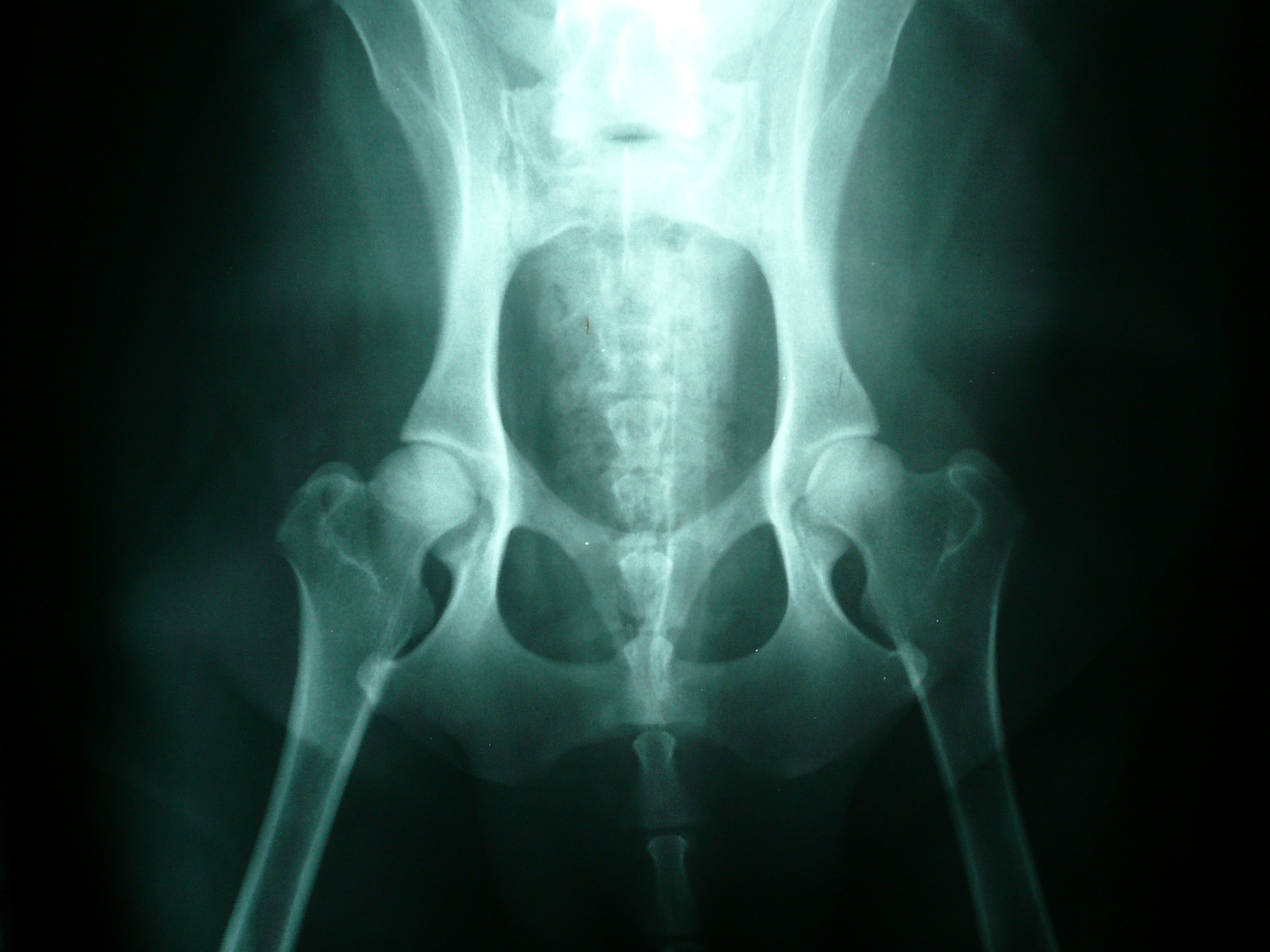
FileNormal canine hips.JPG Wikimedia Commons
You will find a detailed guide on a dog’s hip joint formation in this article. Parts of a dog labeled diagram. The dog's hip is located at the caudal part of the body that extends from the coxal tuber to the ischiatic tuberosity. Ecvdi, utrecht, netherland) were categorized topographically into seven chapters (head, vertebral column,.

A Simple Guide to Hip Dysplasia in Dogs PetHelpful
1) all puppies are born with perfectly normal hips. They have small, tight feet, walking on their toes; Organs and structures of different regions of a dog. The muscles of the rump lie over the lateral and caudal part of the pelvic wall. Dog anatomy details the various structures of canines (e.g. This is why.

anatomy+of+the+thigh+and+hip Pictured above shows the dog muscle
Each os coxae is developmentally composed of the ilium, ischium, pubis, and acetabular bones, which fuse at 12 weeks of age in the dog. The pelvis and hip joint. Regions of the pelvic limb. Features of the anatomical model include normal right coxofemoral joint, left coxofemoral joint showing hip dysplasia and osteoarthritis, disc with herniation.

Canine Pelvic Anatomy Anatomical Charts & Posters
The major bones in a dog’s hind legs are listed below, from the top of the leg to the paw: The canine hip model is a proportional model of the pelvis, sacrum, and lower vertebra, with articulating femurs. Main bone in lower leg. Again, the hip joint is located between the articular. Hip dysplasia is.

Canine Pelvis X Ray
It's much easier to understand how the hip works and how things can go awry with the development of hip dysplasia if you can visualize the anatomy. Main bone in lower leg. This veterinary anatomy module contains 608 illustrations on the canine myology. That is, they are normal for. The condyles are oriented near the.

Canine Hip Model 9060 Dog Pelvis Anatomy GPI Anatomicals
The front legs are loose and flexible, with only muscle attaching them to the torso. Here are presented scientific illustrations of the canine muscles and skeleton from different anatomical standard views (lateral, medial, cranial, caudal, dorsal, ventral / palmar.). Small bone beside the tibia. Ecvdi, utrecht, netherland) were categorized topographically into seven chapters (head, vertebral.

Dog Hip Anatomy Bones, Muscles, and Vessels (Canine Hip Joint Anatomy
The condyles are oriented near the transverse plane to allow cervical spine rotation. Small bone beside the tibia. What is canine hip dysplasia? This is a really nice video tour of the hip and pelvis, and even though it talks about the human hip, the anatomy is. That is, they are normal for. Main bone.

Canine Pelvis Hip Anatomical Model
Light photomicrographs of normal ( g) and fibrillated ( h, arrow) articular cartilage. That is, they are normal for. You will find a detailed guide on a dog’s hip joint formation in this article. Features of the anatomical model include normal right coxofemoral joint, left coxofemoral joint showing hip dysplasia and osteoarthritis, disc with herniation.

What You Need To Know About Hip And Joint Health For Dogs
Find specific details on this topic and related topics from the msd vet manual. Small bone beside the tibia. Imaios is a company which aims to assist and train human and animal practitioners. Approach to the craniodorsal aspect of the hip joint through a craniolateral incision in the dog. The canine pelvis is composed of.

Dog Pelvis Hip Anatomy Anatomy of a Canine Pelvis Hip — Stock Photo
Hip joints from 43 dogs (bodyweight >20 kg) with an average age of 6 years were investigated. The dog's hip is located at the caudal part of the body that extends from the coxal tuber to the ischiatic tuberosity. Anatomy of canine hip dysplasia. The major bones in a dog’s hind legs are listed below,.
Canine Hip Anatomy The pelvis and hip joint. Frequently asked questions on dog’s body parts. Muscle, organ and skeletal anatomy). What is canine hip dysplasia? Exploring the canine hip joint and its supporting structures empowers your therapeutic perspective, helping you choose the best treatment techniques for the.
Dogs Have Disconnected Shoulder Bones (Lacking The Collar Bone Of The Human Skeleton) That Allow A Greater Stride Length For Running And Leaping.
Exploring the canine hip joint and its supporting structures empowers your therapeutic perspective, helping you choose the best treatment techniques for the. This is a really nice video tour of the hip and pelvis, and even though it talks about the human hip, the anatomy is. Serving healthcare professionals through interactive anatomy atlases, medical imaging, collaborative database of clinical cases, online courses. Each os coxae is developmentally composed of the ilium, ischium, pubis, and acetabular bones, which fuse at 12 weeks of age in the dog.
Here Are Presented Scientific Illustrations Of The Canine Muscles And Skeleton From Different Anatomical Standard Views (Lateral, Medial, Cranial, Caudal, Dorsal, Ventral / Palmar.).
Size is not the only issue, with facial structure, leg length and much more varying greatly between breed. Approach to the ilium through a lateral incision. Connects to the hip bone via the hip joint. Hip dysplasia is a common skeletal condition, often seen in large or giant breed dogs, although it can occur in smaller breeds, as well.
Positional And Directional Terms, General Terminology And Anatomical Orientation Are.
The canine hip model is a proportional model of the pelvis, sacrum, and lower vertebra, with articulating femurs. You will find a detailed guide on a dog’s hip joint formation in this article. Light photomicrographs of normal ( g) and fibrillated ( h, arrow) articular cartilage. Frequently asked questions on dog’s body parts.
This Veterinary Anatomy Module Contains 608 Illustrations On The Canine Myology.
Imaios is a company which aims to assist and train human and animal practitioners. It's much easier to understand how the hip works and how things can go awry with the development of hip dysplasia if you can visualize the anatomy. The condyles are oriented near the transverse plane to allow cervical spine rotation. Multiple studies have demonstrated that all normal puppies are born with perfect hips;