
Buccal Region Anatomy - The buccal space is an area created by the buccinator muscle and buccophayrngeal fascia medially, the cheek skin laterally, the lip muscles anteriorly, the pterygomandibular raphe posteriorly, the zygomatic arch superiorly, and the lower aspect of the mandible inferiorly.
Buccal Region Anatomy - It contains the parotid duct, blood vessels, and branches of the facial nerve, as well as the buccal pad of fat. The oral cavity, or more commonly known as the mouth or buccal cavity, serves as the first portion of the digestive system. The buccal space (also termed the buccinator space) is a potential space in the cheek, and is paired on each side. Anatomy, definitions, functions and innervation of the mouth, tongue, salivary glands and fauces. The buccal region is located just inferior to the infraorbital and zygomatic region, and comprises the inferior portion of the cheek.
It is a potential space in the cheek, and is paired on each side. The normal anatomy of the buccal space. It contains the parotid duct, blood vessels, and branches of the facial nerve, as well as the buccal pad of fat. The buccal space (also termed the buccinator space) is a fascial space of the head and neck (sometimes also termed fascial tissue spaces or tissue spaces). The buccal space is superficial to the buccinator muscle and deep to the platysma muscle and the skin. The buccal space (also termed the buccinator space) is a potential space in the cheek, and is paired on each side. During surgery, the harvesting surgeon must be aware of the structures within this anatomical space.
![Schematic drawing of the oral cavity [97]. Download Scientific Diagram](https://i2.wp.com/www.researchgate.net/profile/Javier-Sotres/publication/259357833/figure/fig5/AS:297123783430169@1447851238813/Schematic-drawing-of-the-oral-cavity-97.png)
Schematic drawing of the oral cavity [97]. Download Scientific Diagram
Figure 1 presents an overview of the anatomy of the buccal space and related spaces. The normal anatomy of the buccal space. The buccal space is superficial to the buccinator muscle and deep to the platysma muscle and the skin. It consists of several different anatomically different aspects that work together effectively and efficiently to.

Dental Articles and Resources
The buccal space (also termed the buccinator space) is a fascial space of the head and neck (sometimes also termed fascial tissue spaces or tissue spaces). Figure 1 presents an overview of the anatomy of the buccal space and related spaces. It consists of several different anatomically different aspects that work together effectively and efficiently.
Nasal Cavity Anatomy Structure, Parts, Supply Kenhub
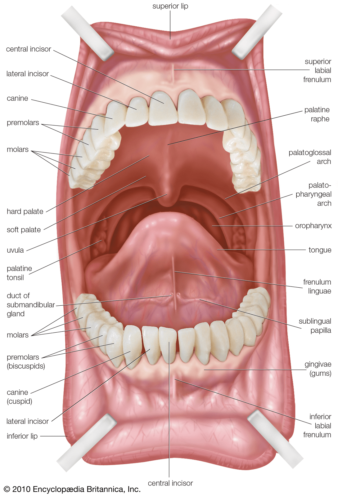
It mainly refers to the area marked by the buccinator muscle. It contains the parotid duct, blood vessels, and branches of the facial nerve, as well as the buccal pad of fat. Fully labeled illustrations and diagrams of the buccal cavity: It consists of several different anatomically different aspects that work together effectively and efficiently.

Oral Cavity Definition, Anatomy, Functions, Diagram
Fully labeled illustrations and diagrams of the buccal cavity: A transverse enhanced ct scan at the level of the upper buccal space shows the lateral projection (open arrow) of buccal fat lateral to the masseter muscle and the medial projection (m) of buccal fat between the masseter muscle and the maxilla. The buccal space (also.

AN3 08 Oral Cavity, Oropharynx, Swallowing StudyBlue
It consists of several different anatomically different aspects that work together effectively and efficiently to perform several functions. The buccal space (also termed the buccinator space) is a potential space in the cheek, and is paired on each side. The buccal space is an area created by the buccinator muscle and buccophayrngeal fascia medially, the.

anatomie de la cavité buccale avec illustration vectorielle. bouche
Figure 1 presents an overview of the anatomy of the buccal space and related spaces. The buccal space is an area created by the buccinator muscle and buccophayrngeal fascia medially, the cheek skin laterally, the lip muscles anteriorly, the pterygomandibular raphe posteriorly, the zygomatic arch superiorly, and the lower aspect of the mandible inferiorly. It.

Pin on Anatomy
The normal anatomy of the buccal space. The oral cavity, or more commonly known as the mouth or buccal cavity, serves as the first portion of the digestive system. It contains the parotid duct, blood vessels, and branches of the facial nerve, as well as the buccal pad of fat. The buccal region is located.
Anatomy of the Oral cavity and salivary glands by Dr Nenad Dordevic
It is a potential space in the cheek, and is paired on each side. The buccal space (also termed the buccinator space) is a fascial space of the head and neck (sometimes also termed fascial tissue spaces or tissue spaces). The buccal region is located just inferior to the infraorbital and zygomatic region, and comprises.

The Oral Cavity Divisions Innervation TeachMeAnatomy
The buccal space (also termed the buccinator space) is a potential space in the cheek, and is paired on each side. Anatomy, definitions, functions and innervation of the mouth, tongue, salivary glands and fauces. A transverse enhanced ct scan at the level of the upper buccal space shows the lateral projection (open arrow) of buccal.

Structure of oral cavity. Human mouth anatomy
It consists of several different anatomically different aspects that work together effectively and efficiently to perform several functions. The buccal region is located just inferior to the infraorbital and zygomatic region, and comprises the inferior portion of the cheek. A transverse enhanced ct scan at the level of the upper buccal space shows the lateral.
Buccal Region Anatomy The buccal space is an area created by the buccinator muscle and buccophayrngeal fascia medially, the cheek skin laterally, the lip muscles anteriorly, the pterygomandibular raphe posteriorly, the zygomatic arch superiorly, and the lower aspect of the mandible inferiorly. The buccal space is superficial to the buccinator muscle and deep to the platysma muscle and the skin. Fully labeled illustrations and diagrams of the buccal cavity: It contains the parotid duct, blood vessels, and branches of the facial nerve, as well as the buccal pad of fat. The buccal space (also termed the buccinator space) is a potential space in the cheek, and is paired on each side.
It Consists Of Several Different Anatomically Different Aspects That Work Together Effectively And Efficiently To Perform Several Functions.
Figure 1 presents an overview of the anatomy of the buccal space and related spaces. The buccal space is an area created by the buccinator muscle and buccophayrngeal fascia medially, the cheek skin laterally, the lip muscles anteriorly, the pterygomandibular raphe posteriorly, the zygomatic arch superiorly, and the lower aspect of the mandible inferiorly. During surgery, the harvesting surgeon must be aware of the structures within this anatomical space. The buccal region is located just inferior to the infraorbital and zygomatic region, and comprises the inferior portion of the cheek.
The Buccal Space Is Superficial To The Buccinator Muscle And Deep To The Platysma Muscle And The Skin.
The buccal space (also termed the buccinator space) is a fascial space of the head and neck (sometimes also termed fascial tissue spaces or tissue spaces). It is a potential space in the cheek, and is paired on each side. The normal anatomy of the buccal space. It contains the parotid duct, blood vessels, and branches of the facial nerve, as well as the buccal pad of fat.
The Oral Cavity, Or More Commonly Known As The Mouth Or Buccal Cavity, Serves As The First Portion Of The Digestive System.
Fully labeled illustrations and diagrams of the buccal cavity: A transverse enhanced ct scan at the level of the upper buccal space shows the lateral projection (open arrow) of buccal fat lateral to the masseter muscle and the medial projection (m) of buccal fat between the masseter muscle and the maxilla. Anatomy, definitions, functions and innervation of the mouth, tongue, salivary glands and fauces. The buccal space (also termed the buccinator space) is a potential space in the cheek, and is paired on each side.
The Buccal Space Is A Region Located In The Cheek, On The Lateral Side Of The Buccinator Muscle.
It mainly refers to the area marked by the buccinator muscle. The buccal space, also known as the buccinator space, is one of the seven suprahyoid deep compartments of the head and neck.