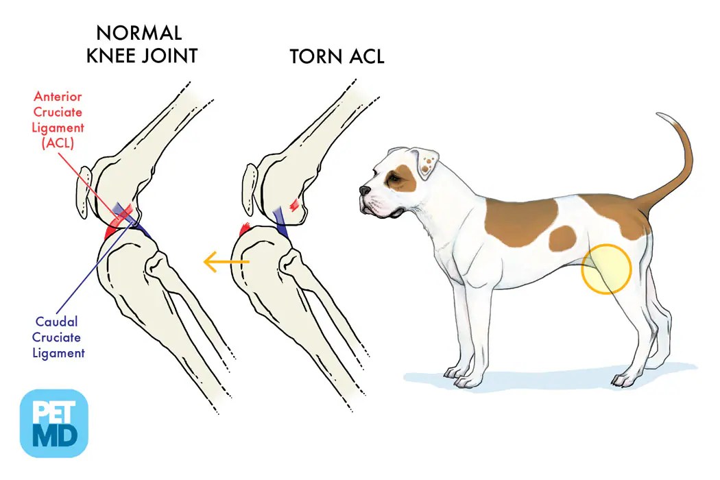
Anatomy Dog Knee Joint - The type of joint formed determines the degree and direction of motion.
Anatomy Dog Knee Joint - The dog stifle (knee) is anatomically very similar to a human knee. The knee joint on a dog is also referred to as the stifle joint. There are two long bones, the femur (thigh bone) and the tibia (shin bone), and a small bone, the patella, which articulate together. It is located on the hind leg, between the femur bone (thigh bone) and the tibia bone (shin bone). It may look like a dog has a continuous thigh, but they have a femur bone as well as.
I will show you the femoropatellar and femorotibial joints separately with their interesting anatomical facts. The hindlimb skeleton of the canine includes the pelvic girdle, consisting of the fused ilium, ischium, and pubis, and the bones of the hindlimb. This diagram shows the most important structures in your knee. The knee is also called the stifle joint, which connects the tibia and fibula with the patella, the dog version of a knee cap. The major bones in a dog’s hind legs are listed below, from the top of the leg to the paw: Muscle, organ and skeletal anatomy). Cranial cruciate ligament (blue/purple), meniscus (red), caudal cruciate ligament (green).
anatomy of the canine knee
The lower thigh (tibia and fibula) is the part of the hind leg beneath the knee to the hock. Examples of them are the joints of bones in the base of the skull, the connection between the body of vertebrae in the spine (drawings later in the article) and the hip (coxae) bones connection (left.
Patellar Luxation (Knee Dislocation) in Dogs Degrees of Severity
A dog’s knee is very much like your knee. The detailing of these structures changes based on dog breed due to the huge variation of size in dog breeds. The major bones in a dog’s hind legs are listed below, from the top of the leg to the paw: Dog anatomy details the various structures.
GPI 9050 Canine Knee Model
This diagram shows the most important structures in your knee. The dog stifle (knee) is anatomically very similar to a human knee. Dog knee and knee cap. The stifle joint is one of the most common orthopedic radiographic studies. Dog leg anatomy is complex, especially dog knees, which are found on the hind legs. You.
Cranial Cruciate Ligament Medical Diagram Torn Knee Ligament in Dogs
Muscle, organ and skeletal anatomy). The dog stifle (knee) is anatomically very similar to a human knee. It may look like a dog has a continuous thigh, but they have a femur bone as well as. Main bone in lower leg. The lower thigh (tibia and fibula) is the part of the hind leg beneath.
Canine Knee Anatomical Model 4Stage Osteoarthritis
Muscle, organ and skeletal anatomy). It is located on the hind leg, between the femur bone (thigh bone) and the tibia bone (shin bone). The upper thigh (femur) is the part of the dog’s leg situated above the knee on the hind leg. Learn about the anatomy of your dog's knee. It may look like.
GPI 9051 Canine 4Stage Knee Model
The lower thigh (tibia and fibula) is the part of the hind leg beneath the knee to the hock. The type of joint formed determines the degree and direction of motion. The knee is also called the stifle joint, which connects the tibia and fibula with the patella, the dog version of a knee cap..
Anatomical ModelCanine Knee
Main bone in lower leg. The insert shows a ruptured cranial cruciate ligament (also note that the shin bone is displaced forward and crushing the meniscus. The dog stifle (knee) is anatomically very similar to a human knee. Dog anatomy details the various structures of canines (e.g. Cranial cruciate ligament (blue/purple), meniscus (red), caudal cruciate.
Anatomy of the canine (dog's) knee joint colorful design, healthy joint
Anatomy atlas of the canine general anatomy: Main bone in lower leg. The knee joint is responsible for providing stability and facilitating movement in the hind leg. The technical term for a dog knee is the stifle joint. Small bone beside the tibia. Technically, the dog knee is on the rear legs. The dog stifle.
ANATOMY OF THE DOG´S KNEE YouTube
The type of joint formed determines the degree and direction of motion. A dog’s knee is very much like your knee. Small bone beside the tibia. Dog anatomy details the various structures of canines (e.g. I will show you the femoropatellar and femorotibial joints separately with their interesting anatomical facts. The dog stifle (knee) is.
Common Knee Problems in Dogs Ortho Dog
Muscle, organ and skeletal anatomy). The detailing of these structures changes based on dog breed due to the huge variation of size in dog breeds. The technical term for a dog knee is the stifle joint. It is located on the hind leg, between the femur bone (thigh bone) and the tibia bone (shin bone)..
Anatomy Dog Knee Joint Illustration of the anatomy of the dog’s knee. The knee is also called the stifle joint, which connects the tibia and fibula with the patella, the dog version of a knee cap. Small bone beside the tibia. This post highlights some of the key elements of the anatomy of the canine knee, and includes information such as why the structures are present, their clinical significance, and associated ligaments. But, let’s see the basic anatomy of the knee joint from the below mentioned labeled diagram.
The Insert Shows A Ruptured Cranial Cruciate Ligament (Also Note That The Shin Bone Is Displaced Forward And Crushing The Meniscus.
Dog anatomy details the various structures of canines (e.g. It is located on the hind leg, between the femur bone (thigh bone) and the tibia bone (shin bone). Fully labeled illustrations and diagrams of the dog (skeleton, bones, muscles, joints, viscera, respiratory system, cardiovascular system). Small bone beside the tibia.
There Are Different Joint Types In The Dog.
Muscle, organ and skeletal anatomy). This post highlights some of the key elements of the anatomy of the canine knee, and includes information such as why the structures are present, their clinical significance, and associated ligaments. The knee joint, or ‘stifle joint’ as it's known in dogs, sits much higher on a dog’s hind legs, and they tend to have short femurs (thigh bones). Cranial cruciate ligament (blue/purple), meniscus (red), caudal cruciate ligament (green).
A Dog’s Knee Is Very Much Like Your Knee.
The detailing of these structures changes based on dog breed due to the huge variation of size in dog breeds. The stifle joint is one of the most common orthopedic radiographic studies. Dog leg anatomy is complex, especially dog knees, which are found on the hind legs. Examples of them are the joints of bones in the base of the skull, the connection between the body of vertebrae in the spine (drawings later in the article) and the hip (coxae) bones connection (left and right in the lower part of the pelvis).
The Dog Stifle (Knee) Is Anatomically Very Similar To A Human Knee.
Connects to the hip bone via the hip joint. Illustration of the anatomy of the dog’s knee. The stifle or knee is the joint that sits on the front of the hind leg in line with the abdomen. Ecvdi, utrecht, netherland) were categorized topographically into seven chapters (head, vertebral column, thoracic limb, pelvic limb, larynx/pharynx, thorax and.


